
ᱞᱮᱣᱟᱴᱳᱨ ᱟᱱᱤ
| Levator ani | |
|---|---|
 Left Levator ani seen from within. | |
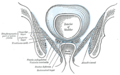
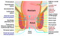
 Coronal section through the male anal canal. B. Cavity of urinary bladder V.D. Vas deferens. S.V. Seminal vesicle. R. Second part of rectum. A.C. Anal canal. L.A. Levator ani. I.S. Internal anal sphincter. E.S External anal sphincter. | |
| ᱯᱟᱥᱱᱟᱣ ᱛᱮ | |
| ᱥᱤᱨᱡᱚᱱ | Inner surface of the side of the lesser pelvis |
| ᱥᱟᱢᱵᱟᱣ | Inner surface of coccyx, levator ani of opposite side, and into structures that penetrate it. |
| Artery | Inferior gluteal artery |
| Nerve | pubococcygeus and iliococcygeus: |
| ᱠᱟᱹᱢᱤ | Supports the viscera in pelvic cavity |
| ᱪᱤᱱᱦᱟᱹᱯᱤᱫ | |
| ᱞᱟᱛᱤᱱ | Musculus levator ani |
| ᱴᱤᱮ | A04.5.04.002 |
| ᱮᱯᱷᱮᱢᱮ | 19087 |
| Anatomical terms of muscle | |

ᱞᱮᱣᱟᱴᱳᱨ ᱟᱱᱤ (levator ani) ᱫᱚ ᱢᱚᱥᱚᱞᱥ ᱜᱩᱴ ᱨᱮᱱᱟᱜ ᱢᱤᱫ ᱵᱽᱨᱚᱰ, ᱯᱟᱛᱞᱟ ᱜᱮ ᱛᱟᱦᱮᱱᱟ, ᱱᱚᱶᱟ ᱫᱚ ᱯᱮᱞᱣᱤᱥ ᱨᱮᱱᱟᱜ ᱟᱭᱤᱫᱚᱨ ᱥᱟᱭᱤᱰ ᱨᱮ ᱢᱮᱱᱟᱜᱼᱟ ᱾ ᱱᱚᱶᱟ ᱞᱮᱣᱚᱴᱳᱨ ᱟᱱᱤ ᱢᱚᱥᱚᱞᱥ ᱨᱮᱱᱟᱜ ᱯᱮᱭᱟ ᱠᱚᱢᱯᱚᱱᱮᱱᱴ ᱨᱮ ᱢᱮᱱᱟᱜᱼᱟ:- ᱯᱭᱩᱵᱳᱠᱳᱥᱤᱡᱮᱭᱟᱥ (the pubococcygeus), ᱤᱞᱤᱭᱚᱠᱳᱥᱤᱡᱤᱭᱚᱥ (the iliococcygeus), ᱟᱨ ᱯᱭᱩᱵᱳᱨᱮᱠᱴᱮᱞᱤᱥ (the puborectalis) ᱠᱚ ᱠᱟᱱᱟ ᱾[᱓]
ᱱᱚᱶᱟ ᱫᱚ ᱵᱷᱤᱛᱨᱤ ᱥᱟᱨᱯᱷᱮᱥ ᱨᱮᱱᱟᱜ ᱥᱟᱱᱟᱢ ᱥᱟᱭᱤᱰ ᱨᱮᱜᱮ ᱯᱮᱞᱣᱤᱥ ᱠᱮᱵᱷᱤᱴᱤ ᱨᱮᱱᱟᱜ ᱮᱴᱮᱪᱰ ᱨᱮ ᱛᱟᱦᱮᱱ ᱠᱟᱱᱟ ᱟᱨ ᱱᱚᱶᱟ ᱫᱚ ᱯᱮᱞᱣᱤᱠ ᱯᱷᱞᱳᱨ ᱨᱮᱱᱟᱜ ᱭᱩᱱᱤᱴ ᱨᱮ ᱟᱹᱰᱤ ᱜᱽᱨᱮᱴᱚᱨ ᱢᱮᱱᱟᱜᱼᱟ ᱾ ᱠᱳᱥᱤᱡᱮᱭᱚᱥ ᱢᱚᱥᱚᱞᱥ ᱫᱚ ᱯᱮᱞᱣᱤᱠ ᱯᱷᱞᱳᱨ ᱛᱮ ᱯᱩᱨᱟᱹᱣ ᱮᱫᱟᱭ, ᱡᱟᱦᱟᱸ ᱫᱚ ᱯᱮᱞᱣᱤᱠ ᱰᱟᱭᱯᱷᱨᱟᱜᱽᱢ (pelvic diaphragm) ᱦᱚᱸ ᱠᱚ ᱢᱮᱛᱟᱜ ᱠᱟᱱᱟ ᱾
ᱱᱚᱶᱟ ᱫᱚ ᱵᱷᱮᱨᱤᱭᱚᱥ ᱥᱴᱨᱚᱠᱪᱟᱨ ᱛᱮ ᱯᱩᱱᱭᱟᱹ ᱥᱮᱫ ᱠᱷᱚᱱ ᱟᱹᱪᱩᱨ ᱟᱠᱟᱱ ᱟᱨ ᱯᱮᱞᱣᱤᱠ ᱠᱮᱵᱷᱤᱴᱤ ᱵᱷᱤᱥᱮᱨᱟᱞ ᱟᱛᱮ ᱜᱚᱲᱚ ᱮᱢ ᱮᱫᱟᱭ ᱡᱟᱦᱟᱸ ᱱᱚᱶᱟ ᱟᱛᱮ ᱜᱮ ᱯᱟᱨᱚᱢ ᱮᱫᱼᱟ ᱾
ᱞᱮᱣᱟᱴᱳᱨ ᱟᱱᱤ ᱫᱚ ᱯᱮᱞᱣᱤᱠ ᱯᱷᱞᱳᱨ ᱢᱚᱥᱚᱞ ᱟᱨ ᱦᱟᱹᱥᱩ ᱠᱟᱱ ᱠᱚᱱᱴᱮᱠᱴ ᱣᱟᱡᱟᱭᱱᱤᱡᱽᱢ ᱚᱠᱛᱮ ᱨᱮᱱᱟᱜ ᱢᱩᱬᱩᱛ ᱠᱟᱱᱟ ᱾ ᱱᱚᱶᱟ ᱫᱚ ᱚᱨᱜᱮᱡᱽᱢ ᱚᱠᱛᱮ ᱠᱚᱱᱴᱮᱠᱴ ᱨᱤᱫᱚᱢᱤᱠᱟᱞᱤ ᱮᱫᱟᱭ ᱾[᱔]
ᱞᱮᱣᱟᱴᱳᱨ ᱟᱱᱤ
[ᱥᱟᱯᱲᱟᱣ | ᱯᱷᱮᱰᱟᱛ ᱥᱟᱯᱲᱟᱣ]ᱞᱮᱣᱟᱴᱳᱨ ᱟᱱᱤ ᱨᱮᱱᱟᱜ ᱯᱮᱭᱟ ᱦᱟᱹᱴᱤᱧ ᱯᱭᱩᱵᱳᱠᱳᱥᱤᱡᱮᱭᱟᱥ(the pubococcygeus), ᱤᱞᱤᱭᱚᱠᱳᱥᱤᱡᱤᱭᱚᱥ(the iliococcygeus), ᱟᱨ ᱯᱭᱩᱵᱳᱨᱮᱠᱴᱮᱞᱤᱥ(the puborectalis) ᱠᱚ ᱠᱟᱱᱟ ᱾
ᱠᱞᱤᱱᱤᱠᱟᱞ ᱥᱤᱜᱽᱱᱤᱯᱷᱤᱠᱮᱱᱴᱥ
[ᱥᱟᱯᱲᱟᱣ | ᱯᱷᱮᱰᱟᱛ ᱥᱟᱯᱲᱟᱣ]ᱞᱮᱣᱟᱴᱳᱨ ᱟᱱᱤ ᱥᱤᱱᱰᱨᱳᱢ ᱫᱚ ᱞᱮᱣᱟᱴᱳᱨ ᱟᱱᱤ ᱢᱚᱥᱚᱞ ᱨᱮᱱᱟᱜ ᱮᱳᱤᱥᱳᱰᱤᱠ ᱨᱮᱠᱴᱟᱞ ᱦᱟᱹᱥᱩ ᱥᱯᱟᱡᱽᱢ ᱦᱚᱛᱮᱛᱮ ᱦᱩᱭᱩᱜ ᱠᱟᱱᱟ ᱾
ᱥᱮᱞᱮᱫ ᱪᱤᱛᱟᱹᱨ ᱠᱚ
[ᱥᱟᱯᱲᱟᱣ | ᱯᱷᱮᱰᱟᱛ ᱥᱟᱯᱲᱟᱣ]-
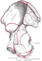 ᱨᱟᱭᱤᱴ ᱦᱤᱯ ᱵᱳᱱ. ᱤᱱᱴᱚᱨᱱᱟᱞ ᱥᱟᱨᱯᱷᱮᱥ ᱾
 -
 Coronal section of pelvis, showing arrangement of fasciæ. Viewed from behind.
 -
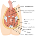 Muscles of male perineum.
 -

-
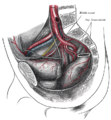 The arteries of the pelvis.
 -
 Sacral plexus of the right side.
 -
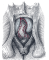
 Iliac colon, sigmoid or pelvic colon, and rectum seen from the front, after removal of pubic bones and bladder.
 -
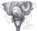
 The posterior aspect of the rectum exposed by removing the lower part of the sacrum and the coccyx.
 -
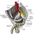
 Male pelvic organs seen from right side.
 -
 Anatomy of the human anus.


ᱥᱟᱹᱠᱷᱭᱟᱹᱛ
[ᱥᱟᱯᱲᱟᱣ | ᱯᱷᱮᱰᱟᱛ ᱥᱟᱯᱲᱟᱣ]- ↑ Essential Clinical Anatomy. K.L. Moore & A.M. Agur. Lippincott, 2nd ed. 2002. Page 217
- ↑ Wallner C, Maas C, Dabhoiwala N, Lamers W, Deruiter M (2006). "Evidence for the innervation of the puborectalis muscle by the levator ani nerve". Neurogastroenterol Motil. 18 (12): 1121–1122. doi:10.1111/j.1365-2982.2006.00846.x. PMID 17109696. S2CID 29088779.
- ↑ Drake, Richard; Vogl, A. Wayne; Mitchell, Adam (2015). Gray's Anatomy for Students (Third ed.). Elsevier.
- ↑ Christopher R. Chapple (2006). Multidisciplinary Management of Female Pelvic Floor Disorders. Churchill Livingstone. pp. 4–. ISBN 978-0-443-07272-7.